New PET tracer improves tumor detection of medullary thyroid cancer tumors and demonstrates potential for targeted therapy.
A newly developed PET imaging agent has been shown to be effective in detecting medullary thyroid cancer (MTC) in preclinical and clinical trials. The results of the study indicate that this PET imaging agent may be a promising therapeutic candidate for clinical use.
The results of the study were published in the Journal of Nuclear Medicine.
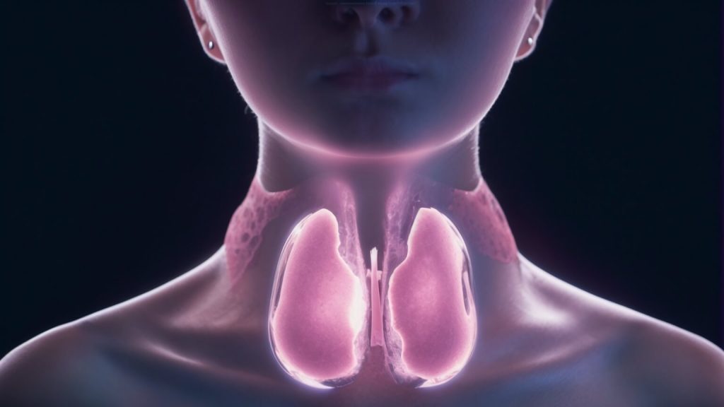
CRC is one of the rarest forms of thyroid cancer, accounting for about 3% of all cases. Because CRC arises from different cells than most thyroid cancers, these patients require different imaging and therapeutic targets.
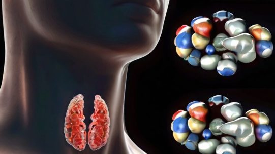
The cholecystokinin-2 receptor (CCK-2R) is overexpressed in most thyroid cancer cells, and various compounds targeting CCK-2R have been developed over the past few years. However, most of these compounds have low metabolic stability, making them less ideal for radioligand therapy. Our team attempted to design several theranostic agents and evaluate their efficacy using simplified design modifications to address the instability issue.”
In the study, three compounds (DOTA-CCK-66, DOTA-CCK 66.2 and external reference DOTA-MGS5) were separately labeled with 64Cu, 67Ga and 177Lu, respectively. The affinity for CCK-2R of each radiolabeled compound was investigated in MTC cells; all three compounds showed high affinity, but DOTA-CCK-66.2 was excluded from further studies due to the better in vitro properties of DOTA-CCK-66.
Stability, biodistribution, imaging and in vivo competition studies were then performed on mice with tumors expressing CCK-2R.68Ga-DOTA-DOTA-CCK-66 was selected for a PET/CT trial based on its overall in vitro and in vivo properties.
Two patients with RRM subsequently underwent PET/CT with 68Ga-DOTA-CCK-66. The compound was well tolerated, showed good biodistribution and high tumor-accumulating activity.
Due to its improved in vivo stability, our compound is well absorbed by tumor and better eliminated from target tissues. This allows for improved lesion detection on PET imaging and further targeted radioligand therapy for MTC,” said Konstantin Lapa, MD, Head of the Department of Nuclear Medicine, University Hospital Augsburg, Augsburg, Germany.
Gunther and Lapa added, “An important result of our study is the idea that pharmacokinetics can be optimized by chemical design. Analyzing the weaknesses of existing compounds and systematically addressing them to improve imaging and therapy is crucial for future clinical applications.”
The study was published online in November 2023.
The authors of the paper are Thomas Günther, Nadine Holzleitner, Roswitha Beck, Hans-Jürgen Wester (Department of Chemistry, Department of Pharmaceutical Radiochemistry, Faculty of Natural Sciences, TUM, Munich University of Technology, Garching, Germany), Oliver Wiering, Georgine Wynand, Alexander Dirks, Christian H. Pfob, Ralf A. Bundschuh, Malte Kircher and Konstantin Lapa (Department of Nuclear Medicine, Faculty of Medicine, University of Augsburg, Augsburg, Germany).